Medical Digital Infrared Thermal Imaging Offers Considerable Benefits
Medical DITI can offer considerable financial savings by avoiding the need for more expensive and invasive investigations.
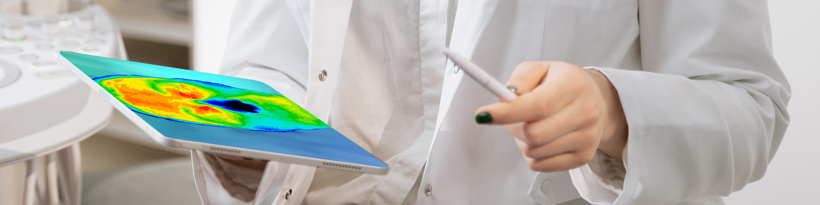
Digital Infrared Thermal Imaging is a non-invasive clinical imaging procedure for assessing a number of disease and physical injuries by showing thermal abnormalities present in the body. Medical Thermography (DITI) enables the examiner to visualize and quantify changes in skin surface temperature. An infrared scanning device is used to convert infrared radiation emitted from the skin surface into electrical impulses that are visualized in color on a monitor. This visual image graphically maps the body temperature and is referred to as a thermogram. The spectrum of colors indicates an increase or decrease in the amount of infrared radiation being emitted from the body surface. Since there is a high degree of thermal symmetry in the normal body, subtle abnormal temperature asymmetries indicating pathology or dysfunction can be easily identified.
Skin blood flow is under the control of the sympathetic nervous system. In healthy individuals, there is a symmetrical dermal pattern which is consistent and reproducible for any individual. This is recorded in precise detail by our medical grade FDA cleared thermography camera, designed and manufactured specifically for medical application with a temperature sensitivity of 0.01 degrees C.
The neuro-thermography application of DITI measures the somatic component of the sympathetic nervous system by assessing dermal blood flow. The sympathetic nervous system is stimulated at the same anatomical location as its sensory counterpart and produces a somato sympathetic response. This response appears on DITI as a localized area of altered temperature with specific features for each anatomical lesion.
Pathology is generally an inflammatory process, i.e. synovitis of joints and tendon sheaths, epicondylitis, capsular and muscle injuries, etc. However, nerve injuries or vascular conditions such a Raynaud’s Syndrome, limb ischemia and DVT may create hypothermic or old patterns. These hot and cold responses may co-exist if the pain associated with an inflammatory focus excites an increase in sympathetic activity.
We can help you plan for better breast health by establishing a baseline early on of your normal thermal patterns so that any changes, no matter how subtle and be readily and easily detected years before self-breast examination and mammography alone. This early detection of thermal asymmetries and thermal abnormalities allows you and your physician to work out the best treatment plan for you. Thermography is not a replacement for structural testing (Mammogram, Ultrasound, MRI) and must be used in conjunction with theses modalities as part of your breast health regime.
Preventative medicine is not limited to breast disease alone. Routine baseline imaging can also screen for arterial inflammation, disc disease, stroke and digestive disorders.
All scans are performed by Certified Clinical Thermographers and all results are interpreted and report by Board Certified MD Thermologists.
Southwest Medical Thermal Imaging, LLC has been Southwest Florida’s premier medical thermography center since 2011. We provide a professional medical thermal imaging service to those wishing to be proactive in their own health and well-being. Referrals are also accepted from physicians, chiropractors, dentists and other health care professionals.